Figure 2.

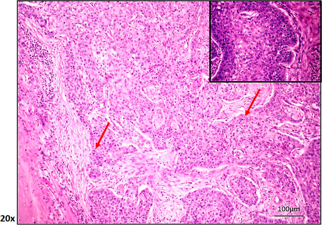
Representative pathological image of a patient with LSCC diagnosis showing invasiveness, heterogeneity, and amorphous cell formation (100 μm hematoxylin and eosin staining; by ImageScope). Red arrow: infiltrative cancer cells.

Representative pathological image of a patient with LSCC diagnosis showing invasiveness, heterogeneity, and amorphous cell formation (100 μm hematoxylin and eosin staining; by ImageScope). Red arrow: infiltrative cancer cells.