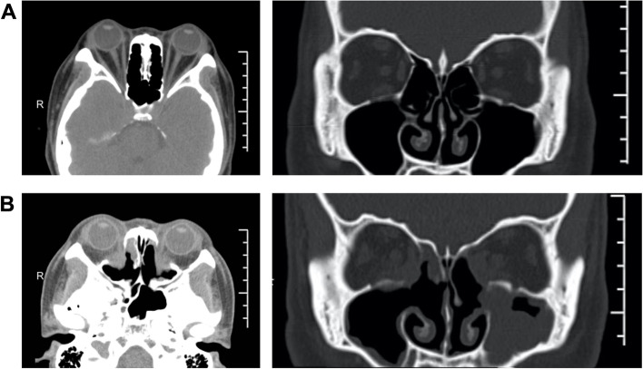
Fig. 5.
CT imaging before and after surgery. A Horizontal and coronal CT images before surgery. CT scans revealed binocular protrusion, increased orbital adipose tissue, and crowding of the orbital apex. B Horizontal and coronal CT images after surgery. A postoperative orbital CT revealed that the exophthalmos had significantly receded, most of the bone in the medial orbital wall's posterior part had vanished, the middle and posterior parts of the orbital cavity had enlarged, the fat herniation was uniform, and the extraocular muscle was not incarcerated

