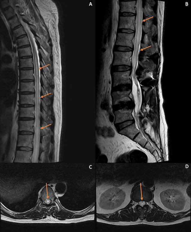
Figure 1.

Sagittal T2-weighted MRI of the thoracic (A) and lumbosacral spine (B) after orthopaedic surgery. Axial T2-weighted images at the T7 (C) and T12 levels (D). A longitudinally extensive hyperintense signal in the intramedullary region of the spinal cord from the T5 level down to the conus medullaris was seen, associated with cord expansion. Axial images show involvement of both sides of the cord
