Figure 2.

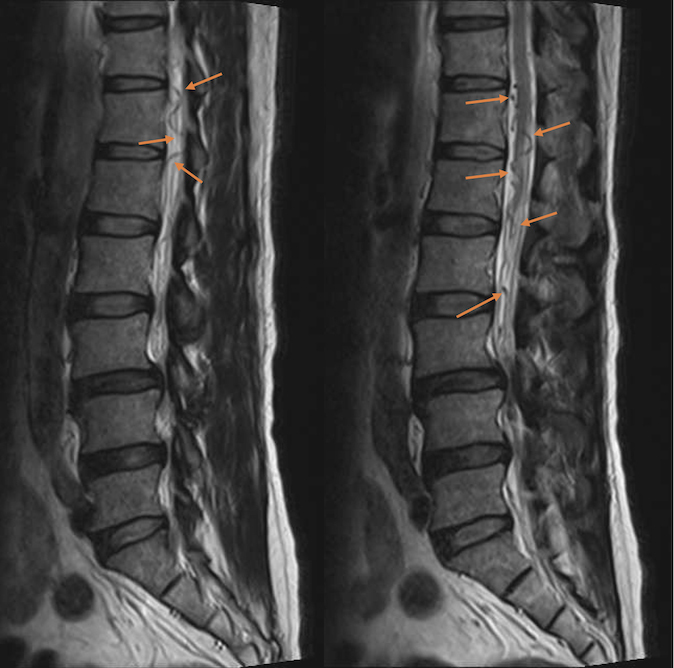
Sagittal T2-weighted MRI of the lumbosacral spine before orthopaedic surgery. Note the prominent, serpentine, tortuous, hypointense lesions around the lower spinal cord and cauda equina, representing dilated perimedullary veins

Sagittal T2-weighted MRI of the lumbosacral spine before orthopaedic surgery. Note the prominent, serpentine, tortuous, hypointense lesions around the lower spinal cord and cauda equina, representing dilated perimedullary veins