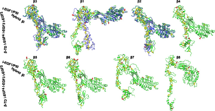
Figure 3. AlphaFold2 structures of 8 human β integrins.
AlphaFold2 structures are depicted in green, while experimentally determined structures for β3 (PDB 3FCS), β1 (PDB 7NXD), and β2 (PDB 4NEH) are shown in blue. The putative N-linked glycosylation sites are represented as red sticks, and disulfide bonds are shown as yellow sticks. The structures were aligned based on the β3 βI domain and are oriented perpendicularly to the cell membrane.

