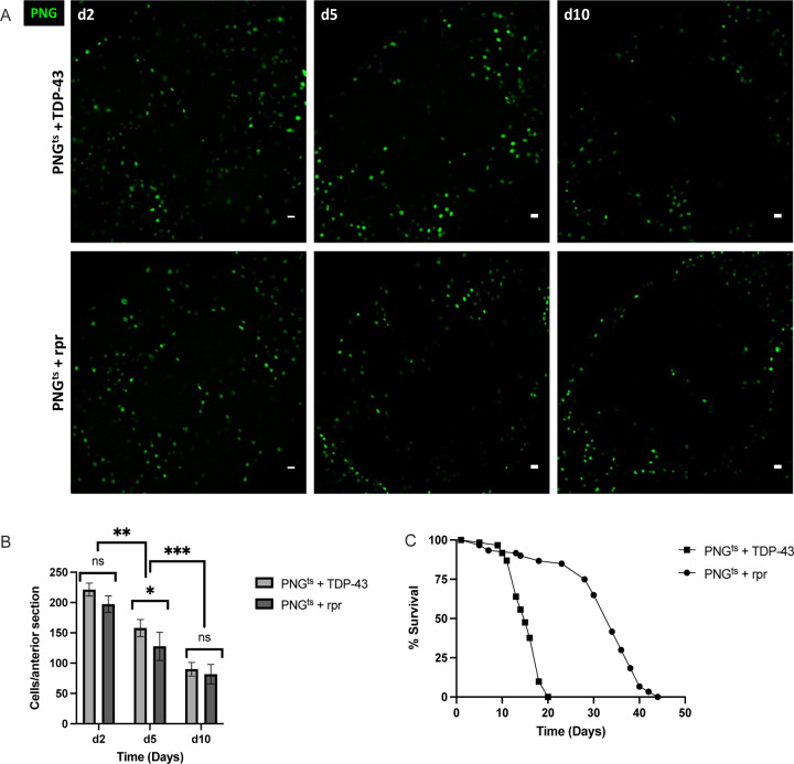
Figure 3. Lifespan deficit observed with TDP-43 expression in PNG is not recapitulated by PNG ablation.
(A) confocal images of an anterior section of the Drosophila brain on days 2,5, and 10 for flies expressing of PNGts + TDP-43 (top) or PNGts + rpr (bottom). (B) quantification of PNG cells in equivalent anterior sections was performed on days 2,5, and 10 post induction by counting the number of nuclei labeled with GFP, which was co-expressed using the UAS-WM reporter (Methods). (C) Lifespan analysis of flies expressing rpr (PNGts + rpr) or TDP-43 (PNGts + TDP-43). Gal4 lines and full genotypes are listed in methods. Scale bar = 10μm. * p<0.5, **p<0.01, ***p<0.001

