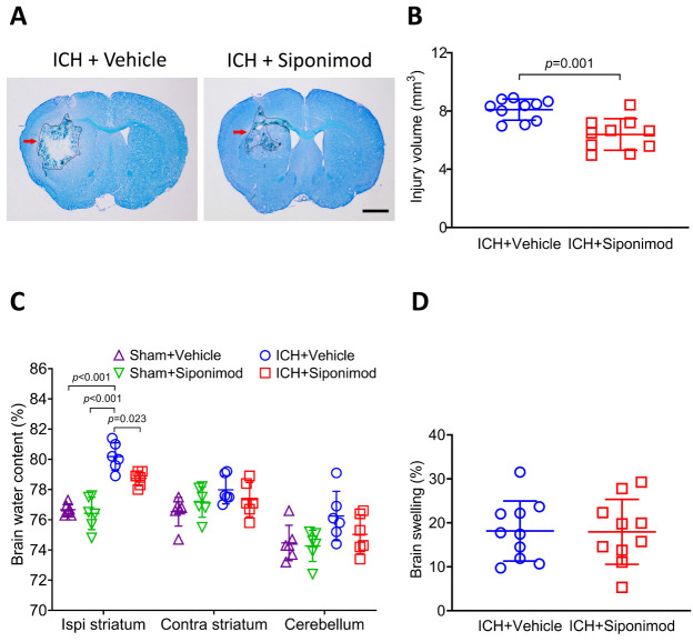
Figure 1.
Siponimod treatment decreased brain injury volume and brain edema on day 3 after ICH. ICH was induced by injecting collagenase into the left striatum of C57BL/6 mice. (A) Representative brain sections were stained with LFB/CV on day 3. The areas of the lesion lacking staining are circled with a black curve (red arrow indicated); scale bar = 1 mm. (B) Brain injury volume was measured in LFB/CV-stained brain sections. The analysis revealed that siponimod treatment decreased brain injury volume compared to the vehicle-treated group on day 3 post-ICH. The t-test for the analysis of brain injury volume. n = 10 mice per group. (C) On day 3 after ICH, siponimod treatment decreased the water content of the ipsilateral striatum. One-way ANOVA followed by Bonferroni’s post hoc test for the analysis of brain water content. n = 6 mice per group. (D) Siponimod treatment did not influence brain swelling on day 3 after ICH. The t-test for the analysis of brain swelling. n = 10 mice per group. All data are expressed as mean ± SD. Ipsi: ipsilateral, Contra: contralateral.

