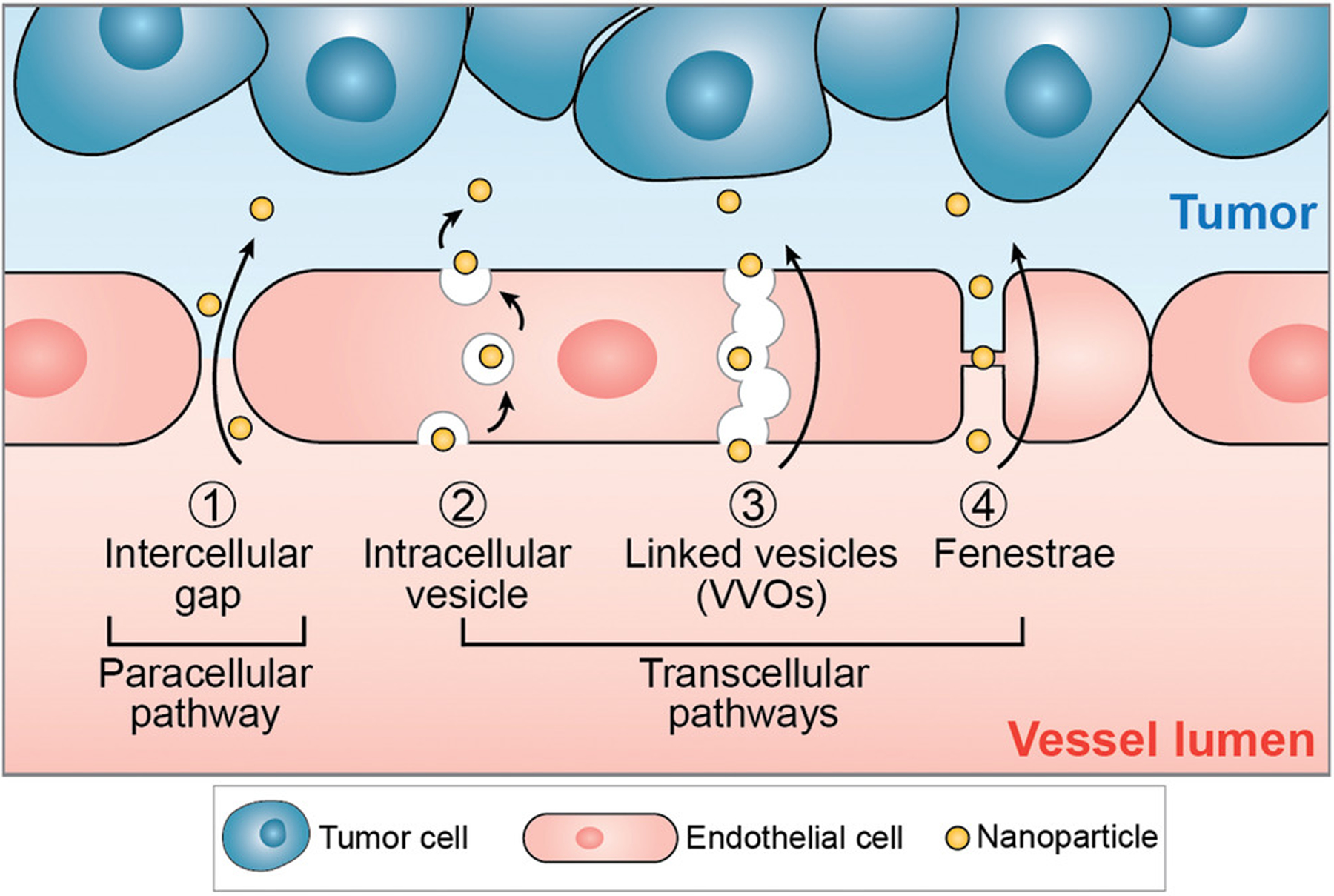
Figure 3.

Nanoparticles can extravasate from tumor vascular lumen into the tumor microenvironment by both paracellular 1) and 2–4) transcellular pathways. For the paracellular pathway, nanoparticles transport passively through gaps in the endothelium, i.e., between adjacent endothelial cells. These intercellular gaps (up to 2 μm in size) result from the abnormal vessel structures caused by rapid tumor angiogenesis and are fundamental for the enhanced permeability and retention (EPR) effect. For transcellular pathways, nanoparticles get transported actively into the tumor microenvironment via intracellular vesicles or through transcellular pores. 2) When transported by intracellular vesicles, nanoparticles first enter the cell and locate in vesicles through endocytosis, then get transported across the cytoplasm, and finally exit the cell through exocytosis. 3) VVO and 4) fenestrae are both trans-endothelial pathways for nanoparticle transport. While VVOs are intracellular organelles composed of linked vesicles, fenestrae represent transcellular pores spanned by a fenestral diaphragm.
