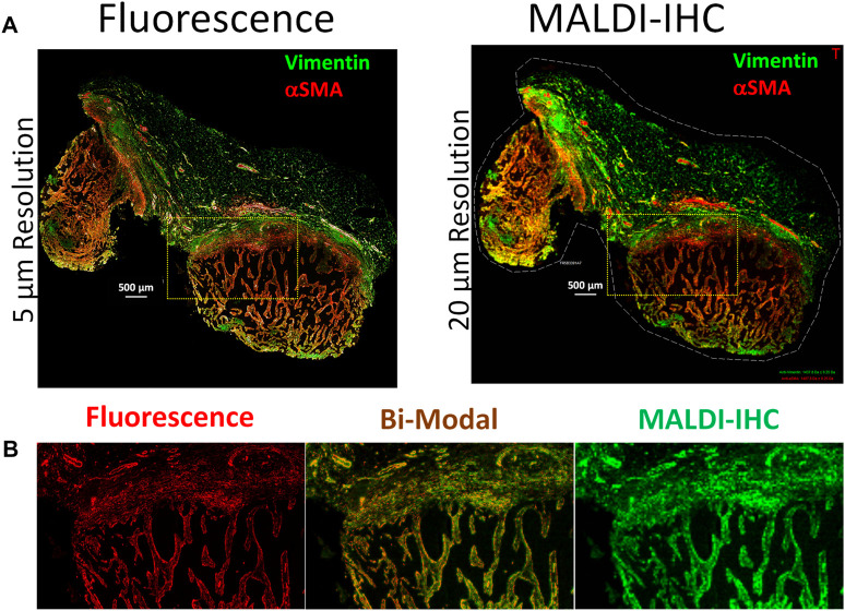
FIGURE 2.
Multimodal imaging of an FFPE breast cancer tissue using dual-labeled Miralys™ probes. (A) Comparison of fluorescence (left) and MALDI-IHC (right) images for the dual-labeled vimentin and alpha smooth muscle actin (αSMA) probes. (B) Magnified region shown from the area within the yellow cross-hatched rectangles in A, for both fluorescence and MALDI-IHC images (left and right, respectively) of the αSMA probe. The center image is a bi-modal interactive merge of both the fluorescence and MALDI-IHC.

