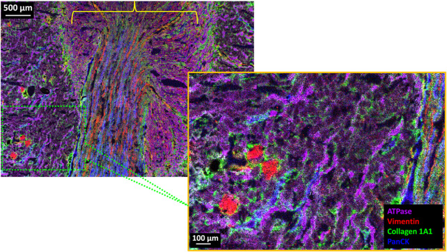
FIGURE 4.
4-Plex MALDI-IHC image of FF normal rat kidney tissue section. (Upper Left) 18 mm2 (5.08 × 3.52 mm) area of the kidney tissue which consists of 715,264 individual pixels which took a total of approximately 11 h of data acquisition (∼18 pixels/s) on a timsTOF fleX equipped with microGRID and recorded at 5 µm pixel size. (Lower Right) 5x magnification of the area shown in the hatched green box which is a portion of the renal cortex. The yellow bracket indicates the general vicinity of the kidney medulla. Base colors for both images are Na+/K+ ATPase (purple), vimentin (red), collagen (green), and panCK (blue).

