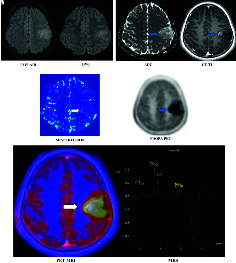
FIG 2.
A 12-year-old boy presented with right focal seizures for a month. Electroencephalography was noncontributory. Imaging-based diagnosis of a tuberculoma was made on the initial contrast-enhanced MR imaging in November 2019. Antitubercular treatment was started empirically. Follow-up MR imaging in February 2022 showed an interval increase of the mass from approximately 1.5 × 1.3 cm to 3.8 × 3.6 cm (images not shown), which led to further work-up, and the patient underwent AA-PET MR imaging and FDOPA-PET MR imaging. T2 FLAIR (A) demonstrates a hyperintense left posterior frontal mass with equivocal diffusion restriction on the diffusion-weighted image (B and C, blue arrow). A predominantly nonenhancing mass with small peripheral nodular enhancement is seen on postcontrast T1-weighted image (D, blue arrow). No apparent increased regional CBV is seen on DSC PWI (E, white arrow). On FDOPA-PET (F, blue arrow) and fused FDOPA-PET MR imaging (G, white arrow), the lesion showed uniformly increased DOPA uptake throughout with a high maximum standard uptake value of 3.62 (lesion/striatum ratio of 1.81 versus <1.0 as normal) and TBR. Multivoxel MRS (H) showed Cho/NAA and Cho/Cr ratios of 2.78 and 1.4, respectively, with a prominent lactate peak. In this patient, FDOPA-PET MR imaging confirmed the precise diagnosis of neoplastic etiology, which, on biopsy, was revealed to be an anaplastic astrocytoma. CE indicates contrast-enhanced.

