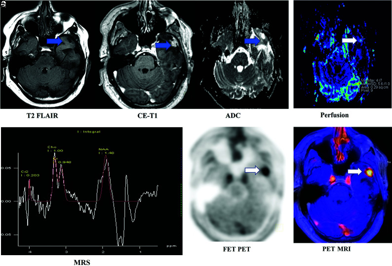
FIG 3.
A 43-year-old man treated for a left anterior temporal lobe glioblastoma (IDH wild-type) status post resection (positive for generalized paroxysmal fast activity, negative for p53 and IDH1, MIB1 labeling index = 10%–12%, fluorescence in site hybridization epidermal growth factor receptor amplification, and no loss of 1p19q) and chemoradiation. He underwent follow-up FET-PET MR imaging after 3 years. T2 FLAIR (A, blue arrow) image shows postsurgical changes in the left anterior temporal lobe with peripheral nodular enhancement on the postcontrast T1-weighted image (B), mild increased diffusion restriction (C), and increased perfusion (rCBV of 6.7). Multivoxel MRS (E) shows noisy spectra with mildly raised choline and an inverted lactate peak. Corresponding increased FET uptake (TBR = 2.8; maximum standard uptake value = 3.7) on FET-PET (F) and fused FET-PET MR imaging (G, white arrow). The patient underwent resection, and pathology showed a recurrent tumor. This case highlights the congruent findings on contrast-enhanced MR imaging and FET-PET with a larger TBR leading to less interobserver variability and improving diagnostic performance for differentiating recurrence from TRC. CE indicates contrast-enhanced.

