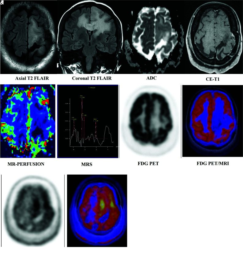
FIG 4.
A 35-year-old man was initially diagnosed with left frontal lobe oligodendroglioma grade II, status post resection and chemoradiation in 2008. He underwent a follow-up [18F] DOPA-PET MR imaging. T2 FLAIR axial (A) and coronal (B) images demonstrate a large cortical and subcortical area of abnormal T2-FLAIR hyperintensity in the left parasagittal frontal lobe extending to the left gangliocapsular area and the corpus callosum and across the midline in the right parietal region without apparent diffusion restriction (C), enhancement (D), and increased rCBV perfusion (E). Multivoxel MRS (F) shows increased Cho/Cr and Cho/NAA ratios (1.86 and 2.31, respectively). FDG-PET MR imaging (G and H) shows no appreciable FDG uptake. FDOPA-PET MR imaging shows areas of significant DOPA tracer uptake (maximum standard uptake value = 1.54 versus <1.0 as normal) more prominently in the left paramedian frontal region. FDOPA-avid, FDG-nonavid nonenhancing lesion in the left frontal region involving the corpus callosum with positive MR imaging correlates suggests active underlying residual/recurrent disease. This case again highlights the superiority of AATs over FDG and the importance of multiparametric MR imaging over individual sequences. AAT uptake in the absence of contrast enhancement and increased perfusion helped with the planning of surgery and radiation. CE indicates contrast-enhanced.

