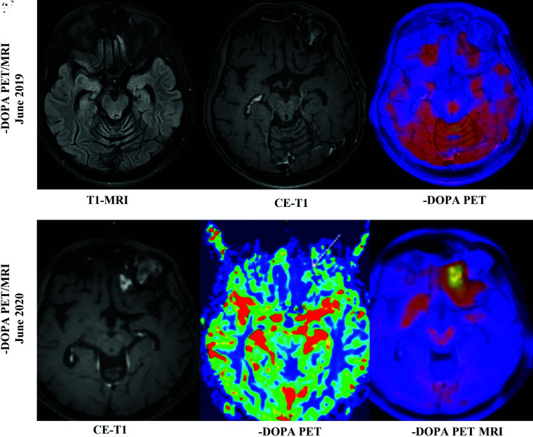
FIG 5.
A 35-year-old man was treated for left frontal glioma (grade II), status post surgical resection and chemoradiation in 2009. He underwent a reoperation in February 2018 for tumor recurrence. In June 2019, [18F] DOPA PET MR imaging (A–C) showed a postresection surgical cavity in the left anterior parasagittal basifrontal region without nodular enhancement or any increased focal FDOPA uptake. There was no evidence of recurrence. In June 2020, follow-up [18F]-DOPA PET MR imaging (D and E) showed a new focal nodular enhancing lesion in the left basifrontal area (arrow) on the postcontrast T1-weighted image (D), with mildly increased rCBV perfusion (E) and corresponding increased FDOPA uptake on fused FET-PET MR imaging (F) (maximum standard uptake value = 3.9; lesion/striatal ratio; 1.38; <1.0 as normal), suggesting recurrence. The patient underwent a reoperation with recurrence found. This case also highlights the congruent findings on contrast-enhanced MR imaging and FDOPA-PET in differentiating TRC from recurrence. CE indicates contrast-enhanced.

