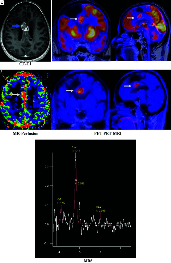
FIG 6.
A 37-year-old man with a known diagnosis of oligodendroglioma, status post resection and chemoradiation. Postcontrast T1-weighted image (A, blue arrow) shows a recurrent enhancing lesion along the inferomedial aspect of the resection cavity of the right frontal region involving the body of the corpus callosum. Fused-PET MR images (B and C) show intralesional increased FDG slightly higher than in the white matter and lower than in the gray matter with increased rCBV perfusion (D, white arrows). Fused FET-PET MR images (E and F) show a relatively larger volume of a recurrent lesion, allowing better estimates of the extent of the lesion. Multivoxel MRS (G) shows an increased choline peak and decreased NAA peak. The pathologic diagnosis was a recurrence. This case highlights the superiority of AAT over FDG, congruent findings on MR imaging and FET-PET, and an excellent TBR, which were helpful in radiation therapy planning and re-surgical resection. CE indicates contrast-enhanced.

