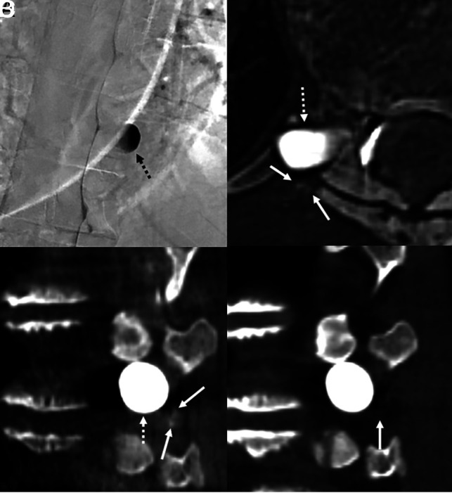
FIG 1.

A 52-year-old woman with years of orthostatic headaches and brain MR imaging demonstrating brain sag and pachymeningeal enhancement. Right lateral decubitus DSM (A) shows a large, right T10 meningeal diverticulum (A, dashed arrow), but no venous opacification was seen on dynamic imaging. Axial (B) and sagittal (C) images from CBCT obtained during contrast injection demonstrate subtle opacification of intramuscular venous branches (B and C, arrows) adjacent to the diverticulum (B and C, dashed arrows), compatible with CSF-venous fistula. Sagittal 50-keV monoenergetic reconstruction from right lateral decubitus CT obtained 15 minutes later (D) no longer shows opacification of these veins in the same location (D, arrow). The patient was treated with transvenous Onyx embolization of the right T10 fistula, with complete resolution of symptoms in 3 months.
