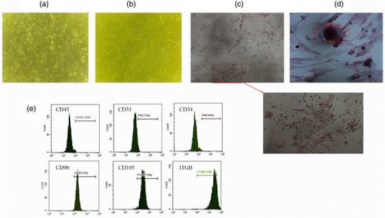
FIGURE 1.

Characterisation and Differentiation of ADMSC. Morphology of canine ADMSCs P0 and P3 was observed under an inverted microscope (100×) (a, b). After 21 days of ADMSC culture in induction medium, lipogenic differentiation induction and osteogenic differentiation induction was performed successfully by staining (100×) (c, d). Markers CD45, CD34 and CD31 exhibited negatively, while CD105, CD90 and ITGB exhibited positively of ADMSC (e).
