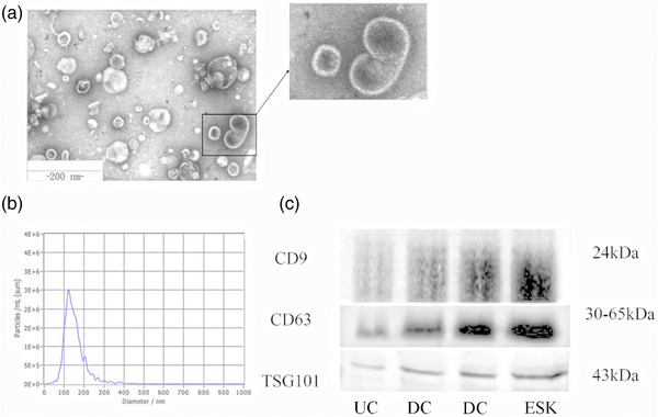
FIGURE 2.

Characterisation of ADMSC‐EVs. ADMSC‐EVs were observed via transmission electron microscopy and characterised as cup‐shape with the diameter around 30–150 nm (a). The particle size distribution of EVs samples was measured by NTA analysis (b). Western blotting against EV markers (CD9, CD63, TSG101) was performed on ADMSC‐EVs (c). EVs were isolated via three methods: UC (ultracentrifugation), ESK (exosome separation Kit) and DC (density gradient centrifugation).
