. 2023 May 16;12:e86413. doi: 10.7554/eLife.86413

Figure 1. Lack of dietary aryl hydrocarbon receptor (AhR) ligands exacerbates cutaneous allergic type 2 responses.

Mice were placed on AhR-poor diet or enriched in indole-3-carbinol (I3C diet) for 3 wk of adaptation prior to the start of experiments. (A–D) Papain or vehicle (PBS) was injected in the footpad at day 0. Mice were sacrificed for analysis either after 24 hr or at day 6. (A, B) Tissues were analyzed by histology 24 hr after papain injection (hematoxylin, eosin, and Safran staining). (A) Representative results (n = 6 per condition). E = epidermis, S = stratum corneum. Bar = 100 μm. (B) Epidermal thickness was measured on images. Median is shown (n = 6 in two independent experiments). One-way ANOVA. (C, D) After 6 d, cells from the draining lymph nodes were analyzed. (C) CD4 T cells and B cells counts. (D) Normalized numbers of T cells were restimulated ex vivo, and cytokine secretion was measured in the supernatant after 24 hr. Median is shown (n = 11–12 in three independent experiments). Kruskal–Wallis test. (E) Papain or vehicle (PBS) was applied topically at day 0. Mice were sacrificed for lymph nodes analysis at day 6. Normalized numbers of T cells were restimulated ex vivo, and cytokine secretion was measured in the supernatant after 24 hr. Median is shown (n = 9 in three independent experiments). Kruskal–Wallis test. For all panels *p<0.05; **p<0.01; ***p<0.001.

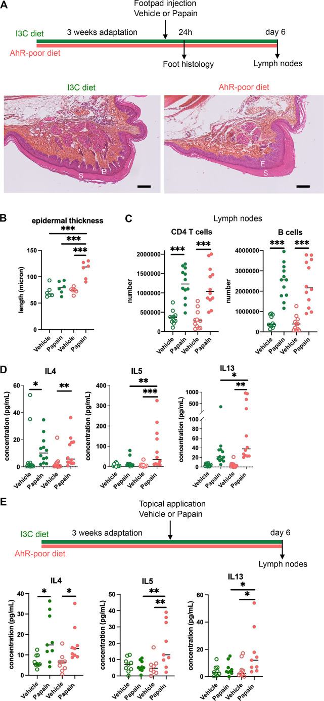
Figure 1.

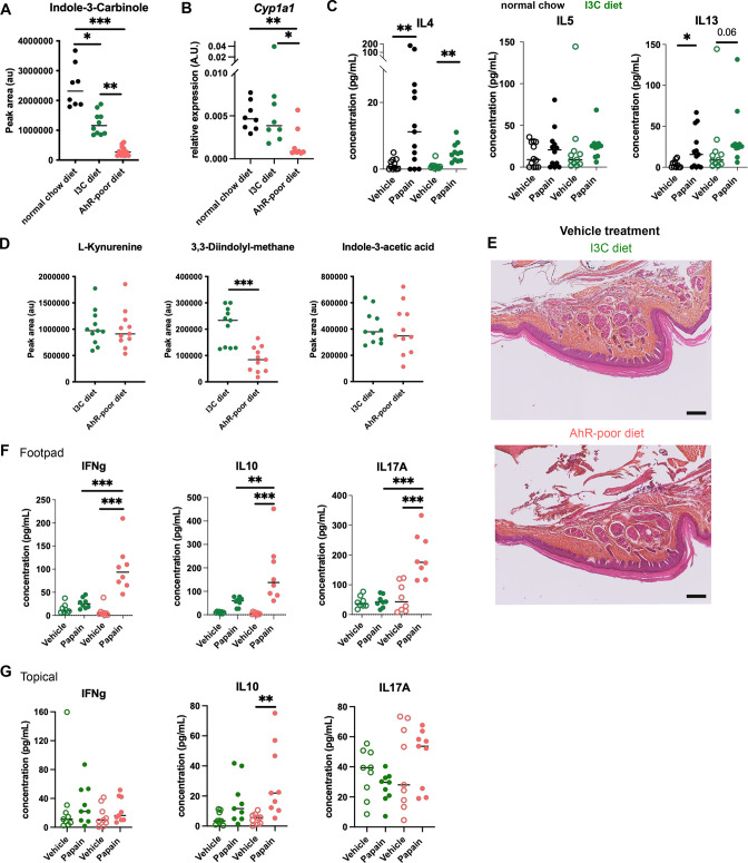
Figure 1—figure supplement 1. Comparable cutaneous allergic Th2 responses in mice fed with normal chow and indole-3-carbinol (I3C) diet.

Figure 1—figure supplement 1.

Mice were placed on aryl hydrocarbon receptor (AhR)-poor or I3C diet for 3 wk of adaptation prior to the start of experiments. (A) Relative abundance of I3C was measured in the serum at day 0. Median is shown (n = 8–11 in two independent experiments). (B) Liver lysates were analyzed by RT-qPCR at day 0. Median is shown (n = 8 in two independent experiments). One-way ANOVA. (C) Papain or vehicle (PBS) was injected in the footpad at day 0. After 6 d, normalized numbers of lymph node T cells were restimulated ex vivo and cytokine secretion was measured in the supernatant. Median is shown (n = 11–13 in three independent experiments). Kruskal–Wallis test. (D) Relative abundance of AhR agonists was measured in the serum at day 0. Median is shown (n = 11 in two independent experiments). (E) 24 hr after vehicle injection, tissues were analyzed by histology (hematoxylin, eosin and Safran staining). Representative results (n = 6 per condition). Bar = 100 μm. (F, G) 6 d after footpad injection (F) or topical application (G), normalized numbers of lymph node T cells were restimulated ex vivo and cytokine secretion was measured in the supernatant. Median is shown (n = 11–12 in three independent experiments). Kruskal–Wallis test. For all panels *p<0.05; **p<0.01; ***p<0.001.