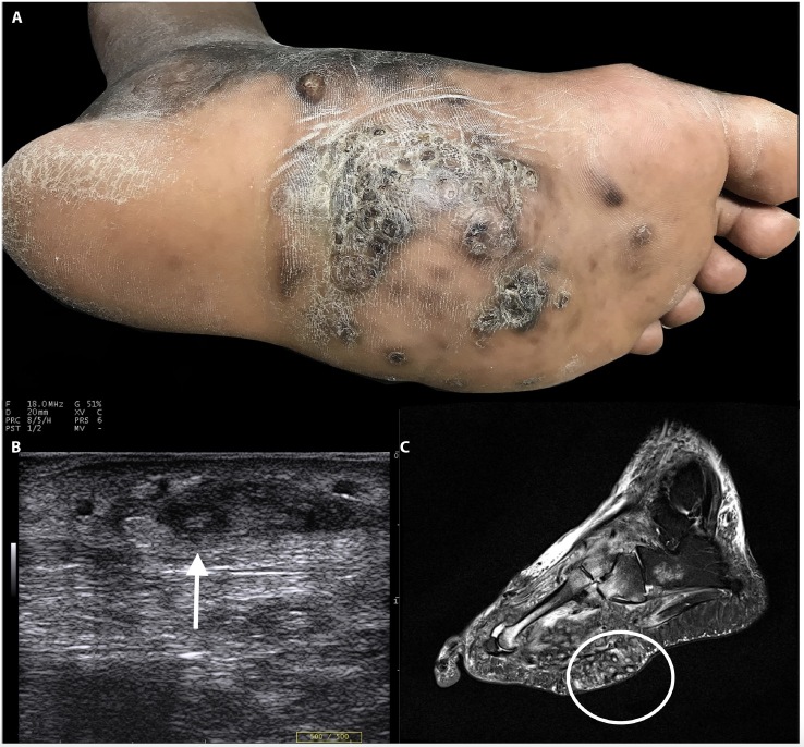
Figure 1.
(A) Painless mass of the plantar surface of the left foot with a purulent discharge. (B) Ultrasound (14–20 MHz Us Transducer – MylabTMOne, Esaote) evidenced a hypoechoic area containing hyperechoic foci, due to granulomatous reaction to bacteria grains. (C) Sagittal T2 weighted STIR MRI confirms the presence of “dots in a circle sign” associated to osteomyelitis.

