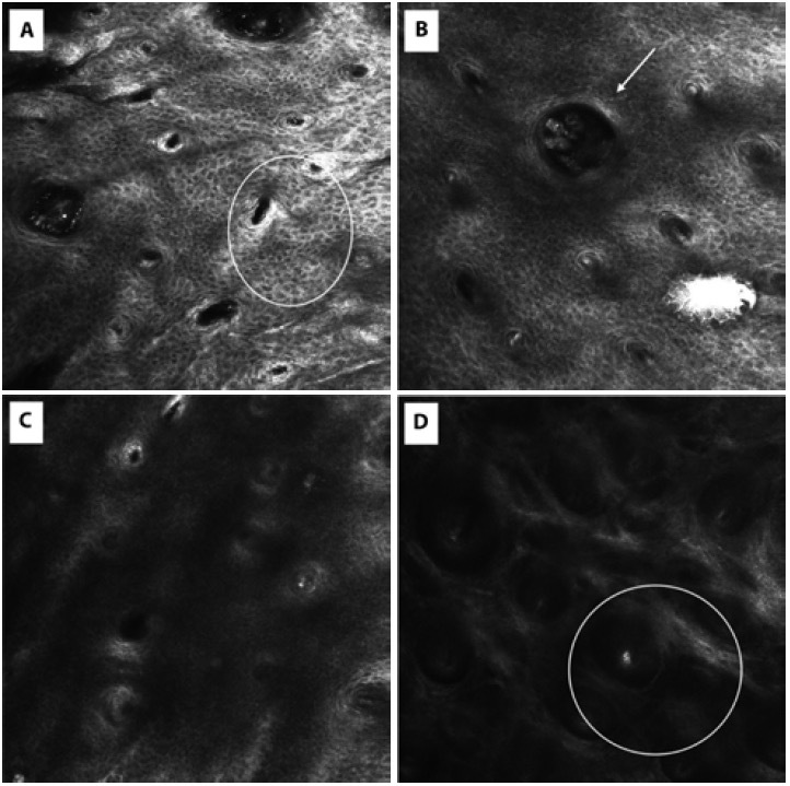
Figure 2.

(A–D) Summary of the main features imaged by in vivo reflectance confocal microscopy (RCM) (Vivascope 3000) in RCM in Inflammatory Linear Verrucous Epidermal Nevus.
(A) Spongiosis (withe circle) with a pattern closer to the honeycombed one with thickened, highly refractive intercellular space between keratinocytes.
(B) Presence of a single roundish refractive Demodex folliculorum (arrow) within the hair follicles and a pattern closer to the honeycombed.
(C) At level of the epithelial-connective tissue junction, non-rimed connective tissue papillae were observed.
(D) Below the basal layer, connective tissue papillae appeared disrupted and confluent in large dark areas (white circle), peculiar signs of the interface dermatitis.
