Figure 1.

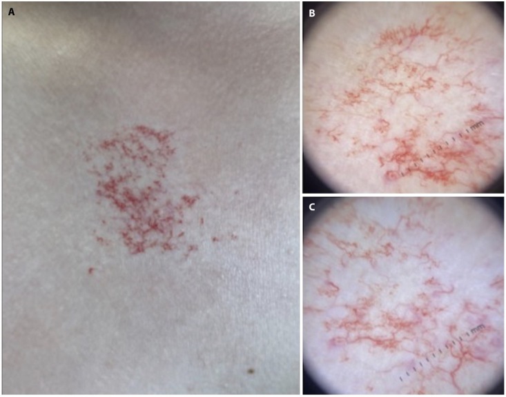
(A) Clinically macule with telangiectasias erythematous background. (B,C) Dermoscopy image showing serpiginous or tortuous vessels, punctate vessels and vascular lacunae.

(A) Clinically macule with telangiectasias erythematous background. (B,C) Dermoscopy image showing serpiginous or tortuous vessels, punctate vessels and vascular lacunae.