Figure 3.

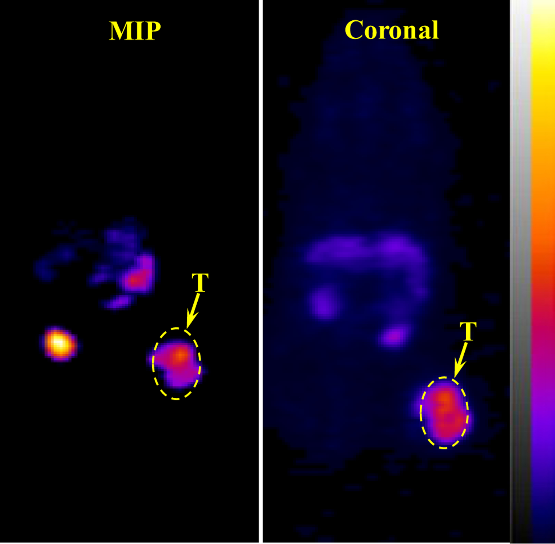
Representative maximum intensity projection (MIP) and coronal PET images of 64Cu-NOTA-PEG2Nle-CycMSHhex on a B16/F10 melanoma-bearing C57 mouse at 2 h post-injection. The melanoma lesions (T) are highlighted with arrows on the images.

Representative maximum intensity projection (MIP) and coronal PET images of 64Cu-NOTA-PEG2Nle-CycMSHhex on a B16/F10 melanoma-bearing C57 mouse at 2 h post-injection. The melanoma lesions (T) are highlighted with arrows on the images.