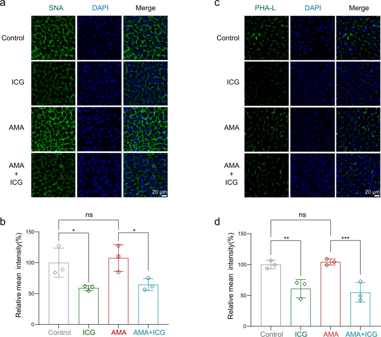
Fig. 8. ICG disrupts glycation in livers in vivo.
The lectin staining assay was used to analyze the glycation of liver sections. The mice were administrated with AMA at 0 h and three consecutive administrations of ICG were injected at 4 h, 6 h, 8 h, and all mice were euthanized at 24 h. The liver sections were stained with SNA for sialylated glycans and PHA-L for complex glycans. a, b Representative images of SNA binding to complex glycans a and semiquantitative evaluation for SNA b by Image J (n = 3 biological replicates). *p = 0.0174, nsp = 0.5946, *p = 0.0136. c, d Representative images of sialylated glycans stained by PHA-L c and semiquantitative evaluation d by Image J (n = 3 biological replicates). **p = 0.0033, nsp = 0.6628, ***p = 0.0008. Data are presented as mean ± SD. The statistics were assessed using a one-way ANOVA test. Source data are provided as a Source Data file.

