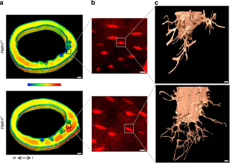
Fig. 8.
FMR1 deletion impacts microstructure of femoral cortical bone and the osteocyte lacuno-canalicular system. a 3D rendering of the femoral mid-diaphysis using µCT imaging, showing localized higher cortical thickness in the lateral region. µCT analysis was implemented to select a ROI (black square) for CLSM. Color bar ranges from 0 µm (blue) to 400 µm (red) thickness. b Z-projection of CLSM images, showing increased canaliculi density in the FMR1-deficient samples. c 3D meshes of lacunae and canaliculi from a randomly selected osteocyte from each ROI FIB-SEM imaging. Scale bars: (a) 100 µm, (b) 10 µm, (c) 1 µm. m medial, l lateral

