Fig. 9.
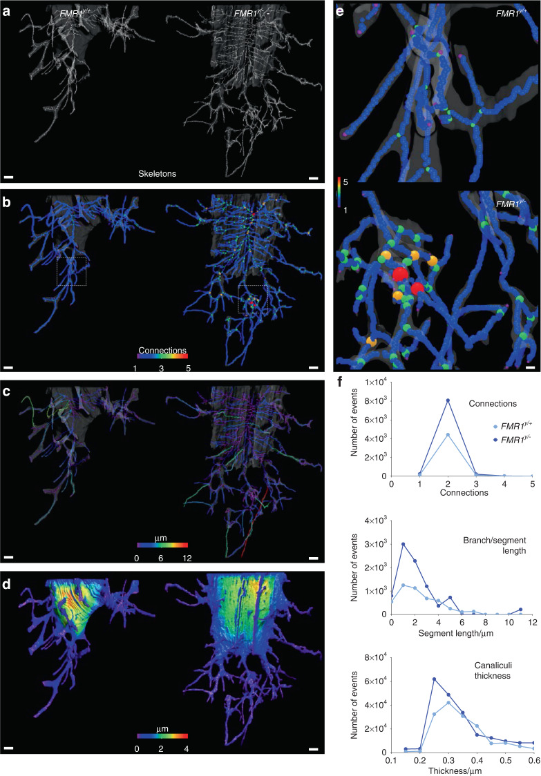
FMR1 deletion impacts the morphology of the osteocyte lacuno-canalicular system at the micro-level. a 3D rendering of skeletons obtained from each volume of interest (VOI) using FIB-SEM. b Connectivity among branches, (c) length of each branch/segment, and (d) overall thickness of the VOI were also 3D rendered. e Close-up view of a region from WT sample (top), where no connections involving 4–5 branches were found, contrasting the close-up view of the FMR1y/− VOI (bottom). Color code indicates number of connections from 1 (blue) to 5 (red). f Number of events was graphed for connections among branches, length of the branches/segments, and regions thinner than 600 nm. Scale bars: (a–d) 1 µm, (e) 200 nm

