. 2023 Apr 6;11(10):3695–3708. doi: 10.1039/d3bm00035d

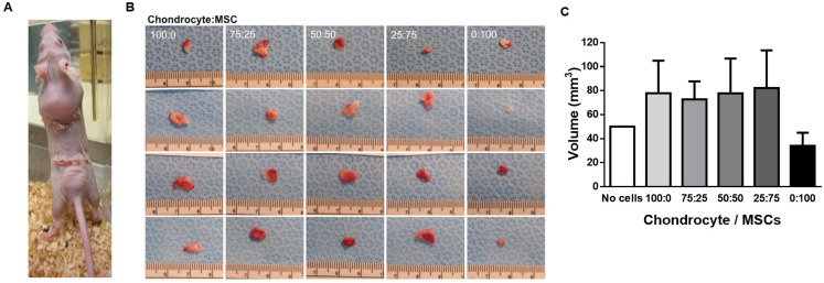
Fig. 6. Effects of subcutaneously implanting PGA scaffolds seeded with co-cultures of isolated rabbit chondrocytes and MSC in nude mice. (A) Anatomical localization and visual size of cell-seeded PGA scaffolds 2 months after implantation (B) Macroscopic aspect and visual size of explants obtained after 2 months of implantation of isolated ear rabbit chondrocytes seeded in PGA scaffolds in co-culture with MSC at different ratios. Ruler is shown below in cm. (C) Quantification of explant sizes obtained after 2 months of implantation of isolated ear rabbit chondrocytes seeded in PGA scaffolds in co-culture with MSC at different ratios. Data are plotted as mean ± SEM.

Fig. 6