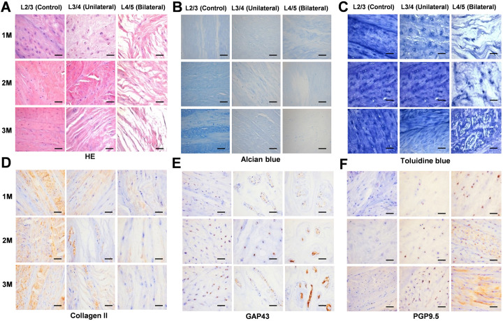
Figure 2.
Histological analysis and immunohistochemistry. A, HE staining showing the structural integrity of the intervertebral disc decreased with increasing ischemia. B, Alcian blue staining indicating that the content of proteoglycan decreases with increasing ischemia. C, Toluidine blue staining indicates that the calcified layer of the intervertebral disc thickens with increasing ischemia. D, With increasing degree of ischemia, the positive expression of collagen II decreased progressively. E, The expression of GAP43 in the intervertebral disc is enhanced with increasing ischemia. F, The expression of PGP9.5 is enhanced with increasing ischemia. The boxed regions are shown at 40 × magnification (scale bar = 100 μm).

