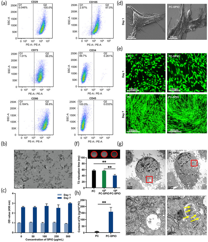
FIGURE 1.

(a) Flow cytometry analysis of the cells isolated from human periodontal ligaments. (b) Transmission electron microscope (TEM) image of superparamagnetic iron oxide (SPIO) nanoparticles. (c) Cell viability of periodontal ligament stem cells (PDLSCs) treated with different concentrations of SPIO nanoparticles. (d) SEM images of PC and SPIO‐labeled PDLSCs (PC‐SPIO) at 1 day (×3000). (e) Live/dead staining of PC and PC‐SPIO at 1 day (×200) and 7 days (×100). (f) In vitro MRI images of different concentrations of PC and PC‐SPIO and their T2 relaxation time. (g) TEM images of PC and PC‐SPIO. SPIO nanoparticles are indicated by yellow arrows. (h) Measurement of the iron content in PC and PC‐SPIO using ICP‐OES. PC represents PDLSCs, and PC‐SPIO represents PDLSCs treated with 250 μg/ml SPIO nanoparticles for 24 h. (n = 3) (*p < 0.05, **p < 0.01)
