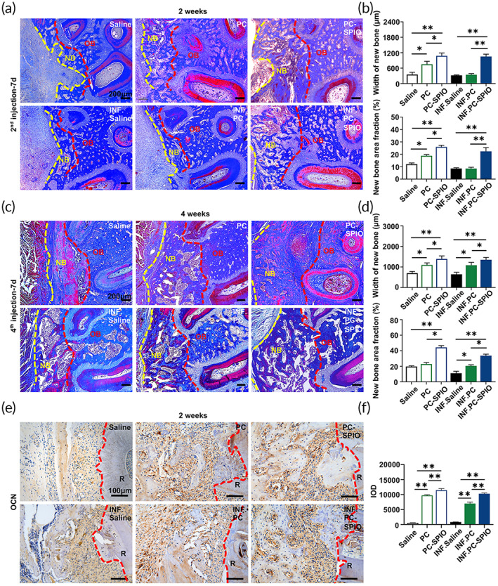
FIGURE 4.

(a) Evaluation of periodontal bone regeneration by Masson staining at 2 weeks after surgery. (b) Quantitative analysis of periodontal bone regeneration by Masson staining, including the width of the buccal new bone and new bone area fraction, at 2 weeks after surgery. (c) Evaluation of periodontal bone regeneration by Masson staining at 4 weeks after surgery. (d) Quantitative analysis of periodontal bone regeneration by Masson staining, including the width of the buccal new bone and new bone area fraction, at 4 weeks after surgery. NB, new bone; OB, original bone. (e) Immunohistochemical staining of osteocalcin (OCN) in the defect area at 2 weeks. (f) Quantitative evaluation of the integral optical density (IOD) of OCN. (n = 3) (*p < 0.05, **p < 0.01)
