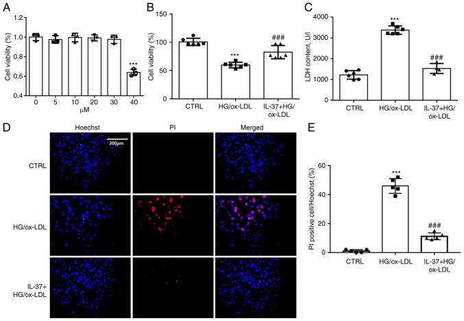
Figure 1.
IL-37 ameliorates the decrease in cell viability caused by HG/ox-LDL. (A) CCK-8 assay, the macrophages were treated with 0, 5, 10, 20, 30 and 40 µM IL-37 for 24 h. The macrophages were divided into three groups: i) CTRL group; ii) HG/ox-LDL group: The macrophages were treated with 100 ng/ml ox-LDL and 25 mM glucose for 24 h; iii) IL-37 group: 30 µM IL-37 for 0.5 h, 100 ng/ml ox-LDL, 25 mM glucose for 24 h. (B) CCK-8 assay. (C) LDH assay. (D and E) Hoechst-PI staining. ***P<0.001 vs. control group and ###P<0.001 vs. HG/ox-LDL. Each dot represents a biological repetition. HG, high glucose; CCK-8, Cell Counting Kit-8; LDH, lactate dehydrogenase.

