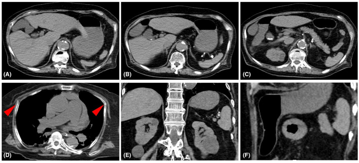
Fig. 1.

Computed tomography scan on admission of a 74‐year‐old woman who underwent cardiac arrest. (A–C) Sequence of images of the abdominal window showing no ascites or splenic injury. (D) Bilateral anterior rib fractures are seen (red arrowheads). (E, F) Coronal and sagittal images of the abdominal window showing no splenic injury.
