
Fig. 2.
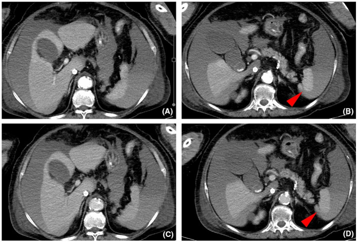
Computed tomography scan with abdominal contrast enhancement of a 74‐year‐old woman who underwent cardiac arrest, on day 4 of admission. (A, B) Arterial phase and (C, D) equilibrium phase. Imaging reveals massive ascites, with an average of 60 Hounsfield units and a suspicious lesion of splenic laceration (B, D, arrowheads) without contrast extravasation.