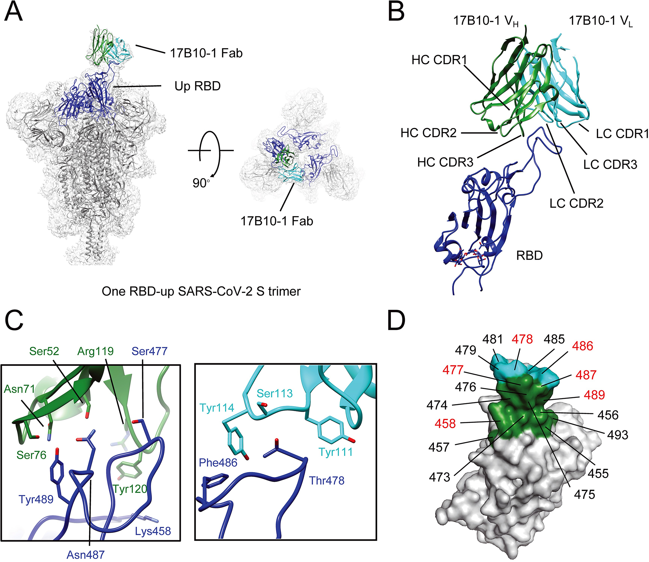
Figure 7.

Cryo-EM structures of 17B10 Fab in complex of a full-length SARS-CoV-2 Spiks (G614) trimer. (A) One 17B10–1 Fab bound to one spike trimer in the one-RBD-up (2.8Å) conformation resolved by Cryo-EM. The EM density is colored in gray, and the structures are represented in ribbon diagram with the RBD in blue and the rest in dark gray. The heavy chain of 17B10–1 is shown in green and the light chain in cyan. (B) Close-up view of the interactions between 17B10–1 Fab and the RBD of the S trimer in the one-RBD-up conformation. (C) Zoom-in views of the binding interface between the heavy (green) or light chain (cyan) of 17B10–1 and the tip of one RBD-up conformation. (D) The footprints on the RBD interacting with the heavy chain (green) or light chain (cyan) of 17B10–1 with the major contacting residues highlighted in red.
