Figure 1.
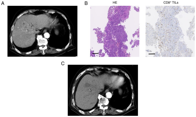
Typical Case 1, high-level CD8+ TILs. (A) CT image of the early arterial phase prior to treatment. (B) HE-staining of liver specimens (magnification, ×100; scale bar, 100 µm). Immunohistochemistry staining for CD8+ T cells was conducted in liver specimens (magnification, ×100; scale bar, 100 µm). (C) CT image of the early arterial phase to evaluate treatment efficacy. TILs, tumor-infiltrating lymphocytes; CT, computed tomography; HE, Hematoxylin and eosin.

