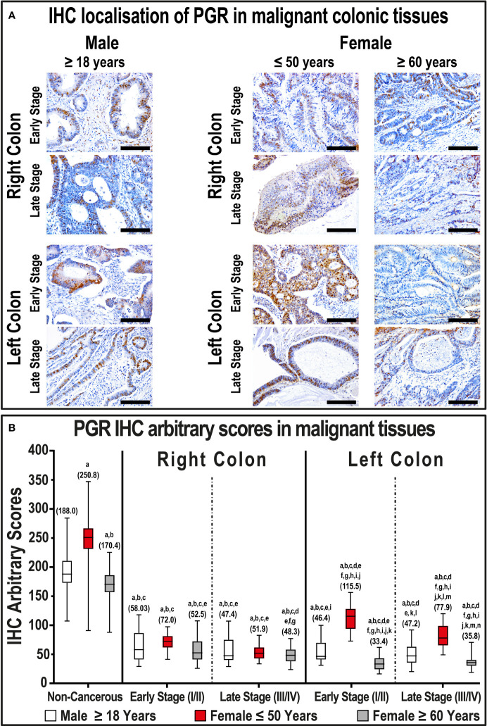
Figure 3.
(A) Immunohistochemical localization of PGR in malignant colonic tissues (n = 120 patients; 20× objective; Scale bar = 15 μm) alongside (B) its IHC arbitrary scores are shown as boxplots according to gender, age, tumor sides and cancer stages. (a = P< 0.05 compared with normal specimens from males ≥ 18 years; b = P< 0.05 compared with normal specimens from females ≤ 50 years; c = P< 0.05 compared with normal specimens from females ≥ 60 years; d = P< 0.05 compared with early-stage right-sided malignant samples from males ≥ 18 years; e = P< 0.05 compared with early-stage right-sided malignant samples from females ≤ 50 years; f = P< 0.05 compared with early-stage right-sided malignant samples from females ≥ 60 years; g = P< 0.05 compared with late-stage right-sided malignant samples from males ≥ 18 years; h = P< 0.05 compared with late-stage right-sided malignant samples from females ≤ 50 years; i = P< 0.05 compared with late-stage right-sided malignant samples from females ≥ 60; j = P< 0.05 compared with early-stage left-sided malignant samples from males ≥ 18 years; k = P< 0.05 compared with early-stage left-sided malignant samples from females ≤ 50 years; l = P< 0.05 compared with early-stage left-sided malignant samples from females ≥ 60 years; m = P< 0.05 compared with late-stage left-sided malignant samples from males ≥ 18 years; n = P< 0.05 compared with late-stage left-sided malignant samples from females ≤ 50 years).

