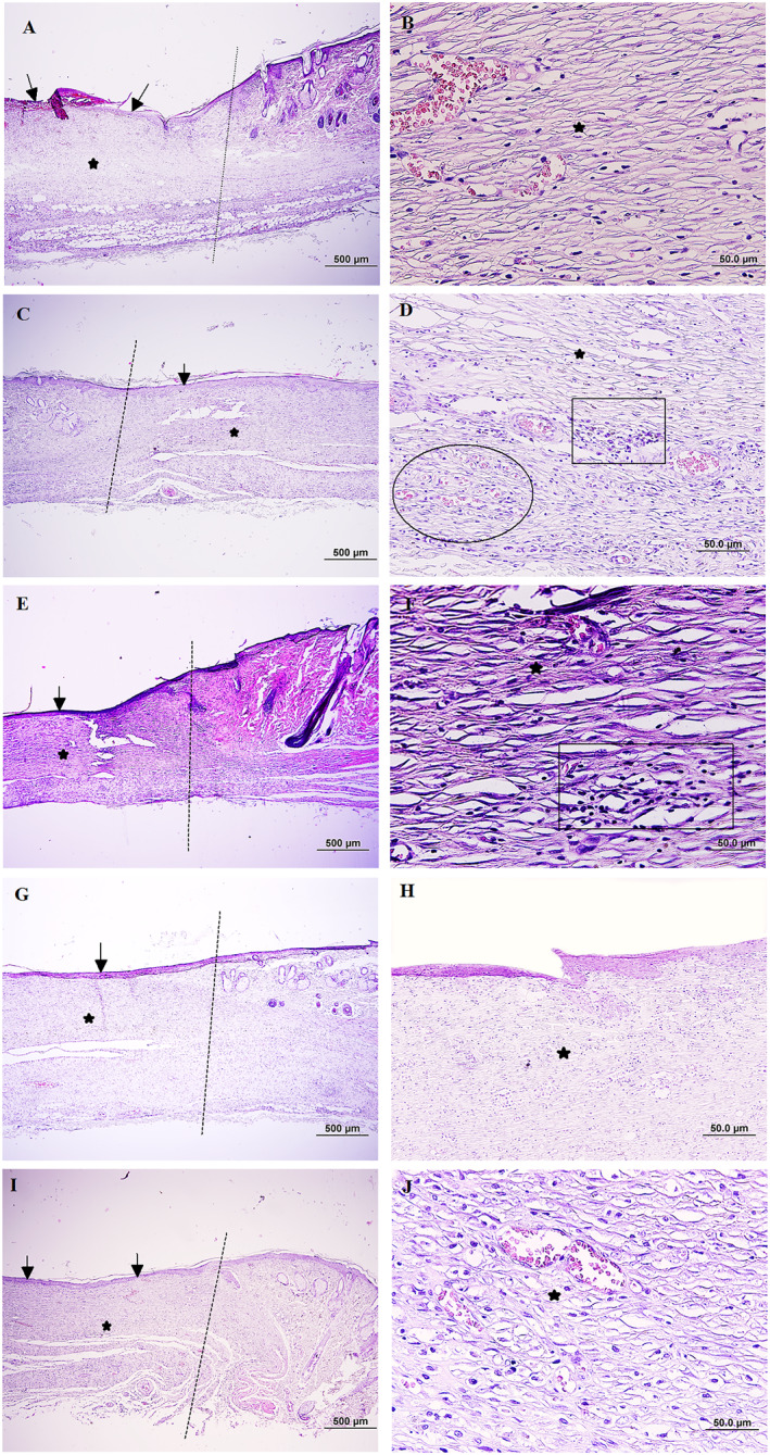
FIGURE 6.

Microscopical evaluation of wound healing 14 days after wound induction, dot lines indicates the demarcation between the intact site and wound site (a): Control group, the partially re‐epithelisation is evident in wound bed (black arrow) and also immature granulation tissue in dermis is shown (black star), (b): Pervious slide with higher magnification, (c) Wound dressing with chitosan, note to appropriate re‐epithelisation (black arrow) and the newly formed dermis accumulated with hypercellular granulation tissue (star), (d): Higher magnification of pervious slide, neoangiogenesis (ellipsoid) and infiltration of inflammatory cells (rectangle) are evident, (e) Wound dressing with epidermal growth factor (EGF), the amount of epithelial regeneration seems appropriate (black arrow), and also note dermal reformation (star) (f) Pervious slide with higher magnification, presence of inflammatory cells in the granulation tissue (rectangle) is seen, (g) Wound dressing with chitosan/EGF nanoparticles ratio 2:1, note to proper re‐epithelisation (black arrow) and the dermis is almost filled with a mature granulation tissue (star), (h): Pervious side with higher magnification, (i): Wound dressing with chitosan/EGF nanoparticles ratio 6:1, the regeneration in epithelium (black arrow) and dermis (star) looks proper, (j) Pervious side with higher magnification, (H & E, Scale bar: A, C, E, G, and (i) 500 μm, B, D, E, H, and (j) 50.0 μm).
