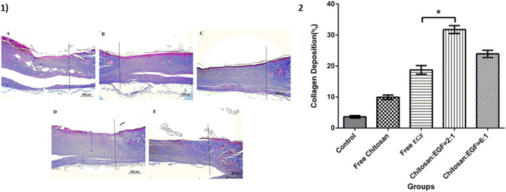
FIGURE 7.

1) Masson's trichrome staining of healed, dot lines indicate the demarcation between the intact site and wound site. Blue staining represents deposition of collagen fibres. (a): Control group, (b): Wound dressing with chitosan, (c): Wound dressing with epidermal growth factor (EGF), (d): Wound dressing with chitosan/EGF nanoparticles ratio 2:1, (e): Wound dressing with chitosan/EGF nanoparticles ratio 6:1, (Masson's trichrome, Scale bar: A‐E: 500 μm). 2) Collagen deposition percentage in different groups after 14 days treatment. Data are represented as mean ± SD (n = 3). *Denotes significant differences (P < 0.0001).
