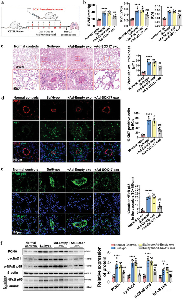
Figure 3.

SOX17‐associated exosomes blocked vascular remodeling, proliferation, and inflammation in mice with Su/hypo‐induced PH. a) The schematic diagram for the construction of PH mice model. b) RVSP, RV/LV+S, and RVH in normal mice, PH mice, and PH mice treated with SOX17‐associated exosomes were examined, ****P < 0.0001 versus Normal controls, ##P < 0.01, ###P < 0.001 versus Su/hypo + Ad‐Empty exo, n = 6–7. c) HE staining of the lung tissues of healthy mice, PH mice, and PH mice treated with SOX17‐associated exosomes, with quantification of vascular wall thickness (80–150 µm), ****P < 0.0001 versus Normal controls, ##P < 0.01 versus Su/hypo + Ad‐Empty exo, n = 6. d) Multiple‐IF stainings for the location and expression of CD31 and Ki67 in the lung tissues of mice were detected, with quantification of percent of Ki67 positive nuclei expression relative to total nuclei in CD31 positive cells, ****P < 0.0001 versus Normal controls, ##P < 0.01 versus Su/hypo + Ad‐Empty exo, n = 6. e) IF staining for the location and expression of NFκB p65 in the lung tissues of mice, quantification of percent of NFκB p65 positive nuclei expression relative to total nuclei in the pulmonary arteries (80–150 µm), ****P < 0.0001 versus Normal controls, ##P < 0.01 versus Su/hypo + Ad‐Empty exo, n = 6. f) Representative western blotting assay for the protein expression of PCNA, cyclin D1, p‐NFκB p65, and NFκB p65 in the lung tissues of mice, **P < 0.01, ****P < 0.0001 versus Normal controls, ###P < 0.001, ####P < 0.0001 versus Su/hypo + Ad‐Empty exo, n = 6–7. Normal cons: Normal control, conditions.
