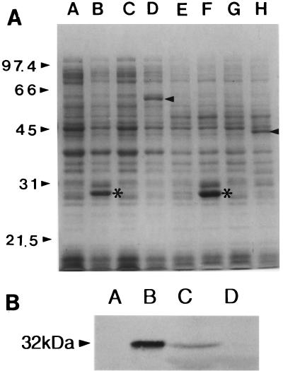
FIG. 6.
Observation of toxRSVv expression by SDS-PAGE (A) and Western blot analysis (B). (A) Expression of ToxRVv and ToxSVv as GST fusion proteins. The molecular masses of ToxR and ToxS were estimated to be 32 and 19 kDa, respectively. Molecular size markers are indicated on the left. In lanes A through H, each pair represents lysates of E. coli DH5α containing corresponding plasmids before and after IPTG induction as follows: A and B, pGEX-3X; C and D, pCMM709; E and F, pGEX-2T; G and H, pCMM820. Arrowheads indicate expressed fusion proteins. Asterisks indicate GST bands expressed after induction. (B) Western blot analysis of ToxRVv expression in V. vulnificus MO6-25/O. Lanes: A, E. coli DH5α harboring pCMM610 without arabinose induction; B, same as lane A except that the cells were induced by 0.2% arabinose; C, V. vulnificus MO6-24/O, the isogenic wild-type strain; D, V. vulnificus CMM981, the toxRVv null mutant. The molecular mass on the left is that of ToxRVv.

