Figure 2.
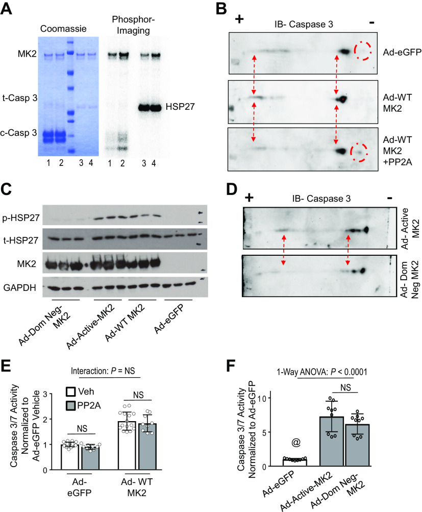
MK2-dependent phosphorylation of caspase-3 does not alter caspase-3 activity. A: kinase activity assays of recombinant activated MK2 (1 μM) and recombinant caspase-3 (100 μM) with 32P-ATP. Coomassie stained gel shows relative protein amounts. Phosphorimaging shows phosphorylation of caspase-3. Lanes—1: 30-min reaction; 2: 60-min reaction; 3: 4–100 nM of HSP27, 30-min reaction as positive control. B: H23 cells were infected with Ad-eGFP or Ad-WT-MK2 and cell lysates underwent two-dimensional immunoblotting for caspase-3. As shown, there is a shift toward the positive electrode on an isoelectric gradient in Ad-MK2 infected H23 cells (red dash arrows). In addition, a band is present toward the negative electrode on an isoelectric gradient in Ad-eGFP infected cells that is not present in Ad-MK2 infected cells (red dash circles). Treatment of cell lysates from Ad-MK2 infected H23 cells with active recombinant serine/threonine phosphatase, PP2A, reverses these charge-based shifts. C: H23 cells were infected with Ad-eGFP, Ad-WT-MK2, Ad-Active-MK2 (constitutively active mutant of MK2) or Ad-Dom Neg-MK2 (a dominant negative mutant of MK2) and 48 h afterwards, cell lysates were analyzed for protein expression. There is significant phosphorylation of HSP27 (the canonical substrate of MK2) in Ad-WT-MK2 and Ad-Active-MK2 but not Ad-eGFP and Ad-Dom Neg-MK2 infected cells. D: H23 cells were infected with Ad-Dom Neg-MK2 or Ad-Active-MK2 and cell lysates underwent two-dimensional immunoblotting for caspase-3. A shift toward the positive electrode on an isoelectric gradient in Ad-Active-MK2 infected H23 cells is not present in Ad-Dom Neg-MK2 infected cells (red dash arrows). E: H23 cells were infected with Ad-eGFP or Ad-WT-MK2 for 48 hours and cell lysates were treated with active recombinant PP2A or vehicle and analyzed for caspase-3/7 activity. There is no interaction between MK2 expression and PP2A treatment on caspase-3/7 activity. There is no within group effect of PP2A treatment on caspase-3/7 activity using post-hoc Tukey’s multiple comparisons test. n = 9–18 per group. F: H23 cells were infected with Ad-eGFP, Ad-Active-MK2 or Ad-Dom Neg-MK2 for 48 h and cell lysates were analyzed for caspase-3 activity. Both MK2 constructs resulted in significant increase caspase-3 activity compared with Ad-eGFP infected cells. There was no difference in caspase-3 activity when comparing Ad-Active-MK2 or Ad-Dom Neg-MK2. @, P < 0.05 vs. all others. All post hoc analyses using Dunn’s multiple comparisons test. n = 9 per group. MK2, mitogen-activated protein kinase-activated protein kinase 2.

