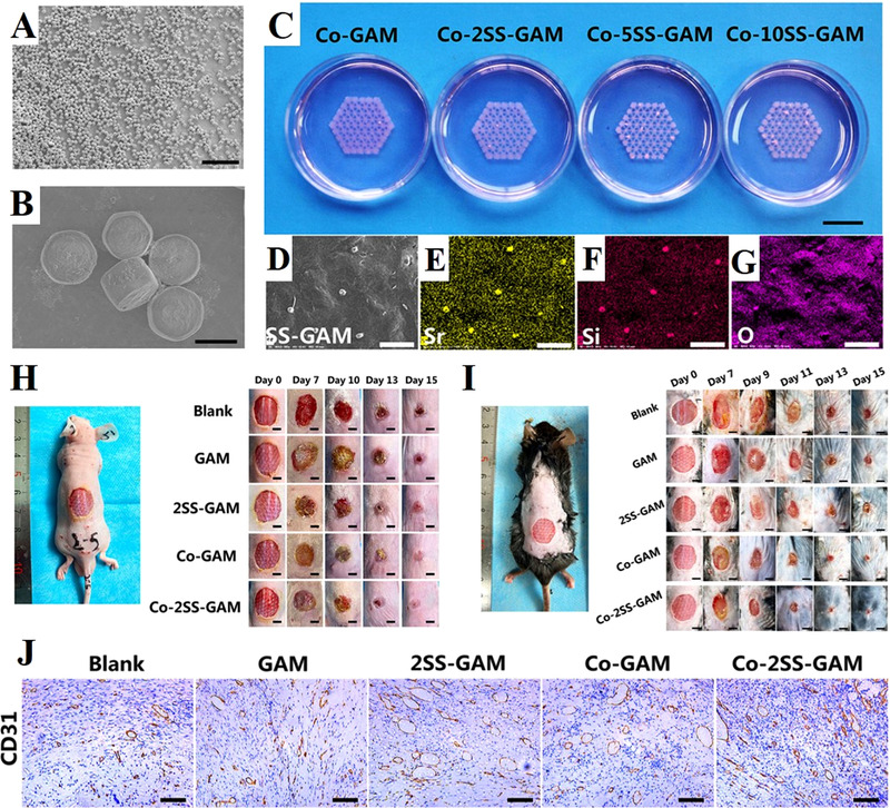
FIGURE 7.

3D bioprinting strontium silicate microparticles‐containing multicellular scaffolds for vascularized skin regeneration. (A,B) SEM images showed the uniform morphology of nearly hexagonal prism of strontium silicate microparticles. (C) Photograph of the 3D bioprinting strontium silicate microparticles‐containing multicellular scaffolds. (D) SEM image of the surface of the scaffold and corresponding EDS elemental mapping of (E) element Sr, (F) element Si, (G) element O. (H) Photos of acute wounds with different treatments at different points of time. (I) Photos of chronic wounds with different treatments at different points of time. (J) Images of immunohistochemical staining with CD31 antibody. The Co‐2SS‐GAM group showed the highest degree of angiogenesis. Reproduced with permission.[ 137 ] Copyright 2021, Wiley‐VCH
