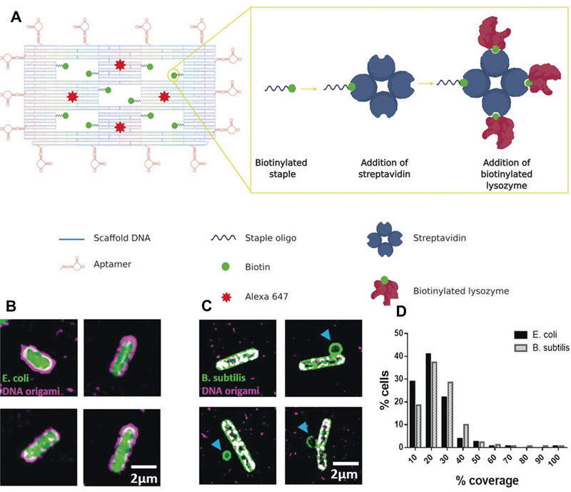
FIGURE 9.

(A) Illustration of DNA origami nanostructure containing five “wells” carrying ten biotinylated staples for streptavidin attachment and fourteen aptamers conjugated with staples for driving the binding with bacteria. (B) Structured illumination microscopy (SIM) imaging of DNA origami (magenta) binding to E. coli (green). (C) SIM imaging of DNA origami (magenta) binding to B. subtilis (green). (D) Mean coverages for E. coli and B. subtilis, respectively. Reproduced with permission from.[ 168 ] Copyright 2020, John Wiley & Sons
