
FIGURE 7.
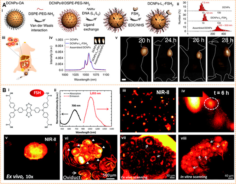
Image of the ovarian tumor on the NIR‐II window. (A) The DCNPs‐L1‐FSHβ nanoprobes accurately targeted ovarian serous carcinoma. Reproduced with permission.[ 180 ] Copyright 2018, Springer Nature. (B) Ovarian follicles granulosa cells of antral/smaller follicles were found in vivo by injecting an FSH‐CH probe. Granulosa cells of secondary follicles (arrows) were also clearly visualized ex vivo and in vitro using a confocal fluorescence microscope. Reproduced with permission.[ 187 ] Copyright 2017, Royal Society of Chemistry.