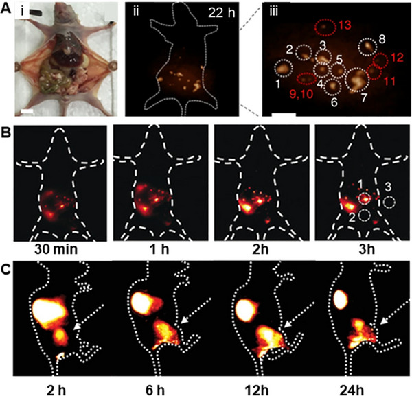
FIGURE 12.

Imaging of distant tumor metastasis. (A) (i) NIR‐II imaging of human ovarian adenocarcinoma peritoneal metastases using DCNPs‐L1‐FSHβ nanoprobe. (ii) Imaging was obtained at 22 h; (iii) the enlargement of the NIR‐II nanoprobes with large peritoneal metastatic tumors (nos. 1–8) and ultra‐small lesions (nos. 9–3). 1000 nm LP filter; Scale bar, 1 cm. Reproduced with permission.[ 180 ] Copyright 2018, Springer Nature. (B) The peritoneal metastasis tumor nodules were lighted by FEAD1. Reproduced with permission.[ 255 ] Copyright 2020, John Wiley & Sons. (C) Lung metastasis from primary osteosarcoma was imaged using CH1055‐PEG. The orthotopic tumor has been indicated by white arrows. Reproduced with permission.[ 257 ] Copyright 2020, John Wiley & Sons.
