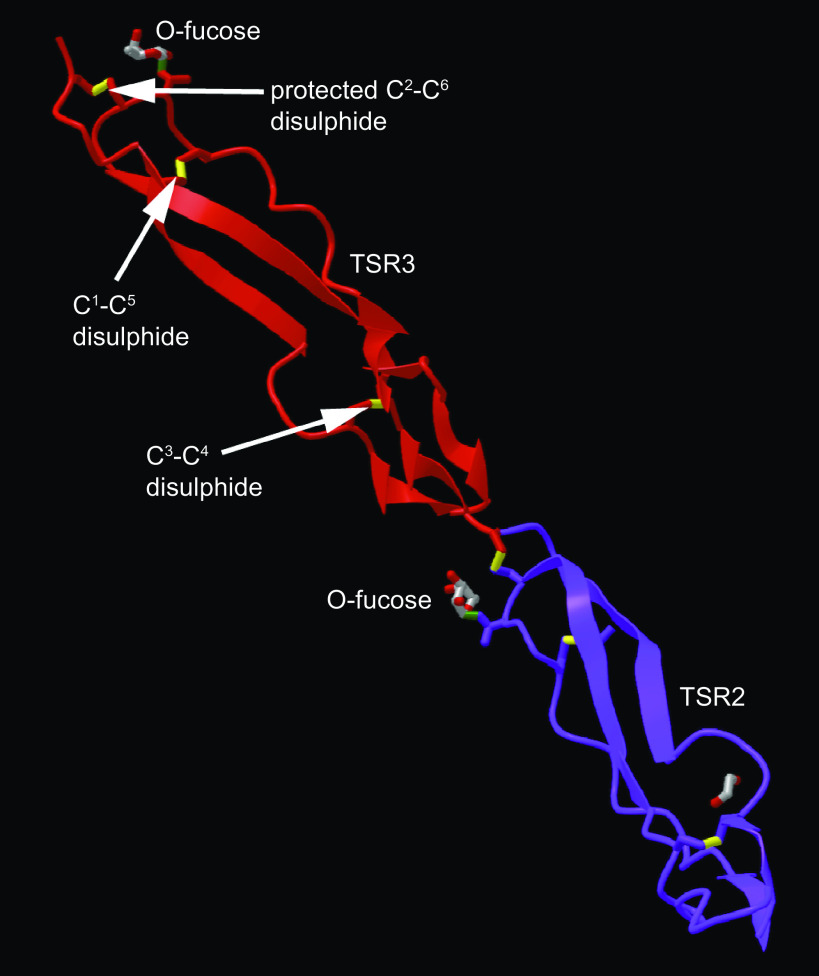
Figure 1.
Posttranslational modifications support stable folding of the TSR domains of thrombospondin-1. The structural model [PDB 7YYK (14)] shows the second (purple) and third (red) TSR domains. Disulfide bonds are in yellow, with cysteines numbered from the N-terminus of the domain. The position of the O-fucose modification that shields a disulfide bond to stabilize the folded domain is indicated in TSR3. Diagram prepared in iCn3D (15) and exported from NCBI. TSR, thrombospondin type 1 domain.

