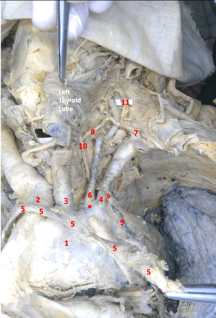
Figure 1. Image of the thoracic region and left neck in gross dissection.
1- Aortic Arch, 2- Brachiocephalic Trunk, 3- Left Common Carotid Artery, 4- Left Subclavian Artery, 5- Left Brachiocephalic Vein (Mobilized), 6- Left Vertebral Artery, 7- Thyrocervical Artery, 8- Left Inferior Thyroid Artery, 9- Thoracic Duct (pulled by the mobilized venous angle), 10- Left Recurrent Laryngeal Nerve, 11- Left Sympathetic Trunk.
*- Direct Origin of the Left Vertebral Artery from the Arch of Aorta (Anomalous Origin)

