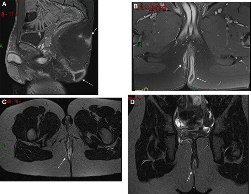
FIGURE 2.

A) Fat suppressed gadolinium enhanced sagittal T1 weighted MRI in a 15-year-old boy with perineal hidradenitis shows a posterior gluteal abscess with sacral subcutaneous oedema. B) Fat suppressed gadolinium enhanced coronal T1 weighted MRI in a 15-year-old boy with perineal hidradenitis shows a posterior bilateral gluteal abscess. C) Fat suppressed axial T2 weighted MRI in a 9-year-old girl with perineal Crohn’s disease show a posterior median fistula. D) Fat suppressed coronal T2 weighted MRI in a 9-year-old girl with perineal Crohn’s disease show a posterior median fistula.
