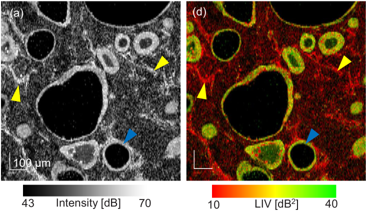
Fig. 2.
(a) En face OCT-intensity and (b) LIV images of a normal alveolar organoid measured 3 days after the organoid establishment. The images were extracted from approximately below the sample surface and the FOV is . The cystic and mesh-like structures are expected to be alveoli and fibroblasts, respectively.

