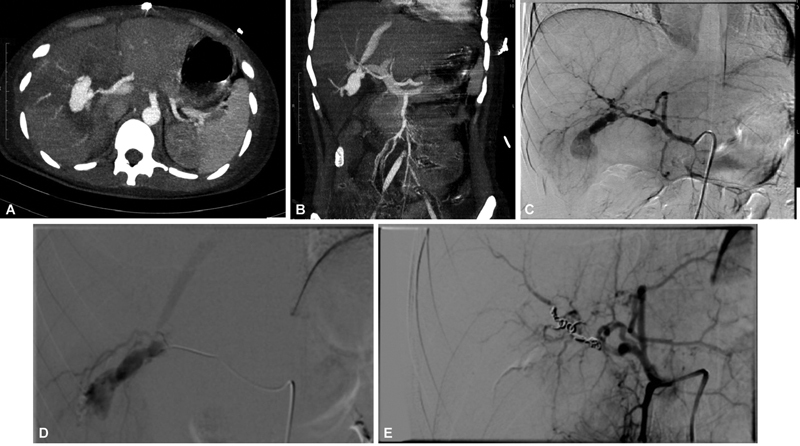
Fig. 2.

( A , B ) Computed tomography (CT) angiogram showing a large right hepatic artery pseudoaneurysm in a patient of firearm injury with shunting into hepatic vein. ( C , D ) Celiac angiogram and right hepatic artery angiogram depicting the pseudoaneurysm and the hepatic venous shunting. ( E ) Post-coil embolization angiogram showing exclusion of aneurysm.
